![]()
Organic chemists make judgements about how to represent molecular structures on a daily basis. When we give lectures, when we write papers, when we scribble on a napkin at lunch to share an idea with a colleague, we must make several choices (whether they be conscious or not). We must decide how much structural information to convey, from what angle to convey it, and whether to use simple lines or elaborate computer-generated graphics. Much has been written on these subjects (some by Roald Hoffmann), so for now I will just present a collection of images that I have produced for use in papers, in lectures, on journal covers, and just for fun.
Other examples of science related artwork can be found at the Chemist's Art Gallery, Molecular Graphics Art Show, Image and Meaning, and Art & Science Collaborations, Inc. websites, as well as David S. Goodsell's, and Nick DeMello's personal websites.
A wonderful book of molecular images that predates the rise of computer graphics is The Architecture of Molecules, by Linus Pauling and Roger Hayward (W. H. Freeman & Co.: San Francisco, 1964).
Some of my non-chemistry images can be found here.
Cube of Cubanes
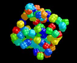
This image was created using a program called AutoDock that I used in my graduate research. It was designed for the automated docking of small ligand molecules into large macromolecular hosts. I discovered (by accident, of course) that if the box of space your ligand is allowed to sample is not centered on the macromolecule, you produce a random distribution of ligand positions inside a cube. I couldn't resist doing this again, this time on purpose and using my favorite cubic molecule. The resulting atomic coordinates were initially visualized with RasMol.
Of Mice
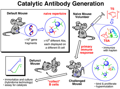
This image was created for use as a slide in several talks I've given on antibody catalysis. It was used to illustrate the key steps in the creation of catalytic antibodies. The image of duplex DNA in the top left panel was actually created from crystallographically determined coordinates using RasMol. The mice pictures are modified versions of an image I found somehwere on the web (unfortunately, I no longer remember where - my apologies to the creater of the original mouse image).
Boat (Full of Chemists) Conquers Chair
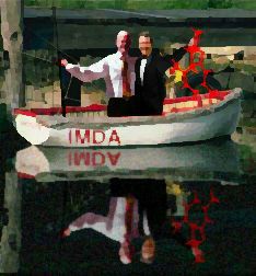
This image was created for the cover of the Journal of Organic Chemistry, but didn't quite make it there. It was to accompany a paper on intramolecular Diels-Alder (IMDA) reactions and was inspired by a call for creativity by my graduate advisor. The "chemists" (stylized versions of two of the paper's authors) are triumphantly hoisting their latest catch: a chairlike transition state for the IMDA reaction of a substituted hexadienyl acrylate.
IMDATSs
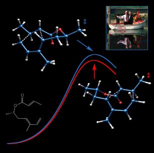
This image is what resulted from the rejection of "Boat (Full of Chemists) Conquers Chair" by the editors of the Journal of Organic Chemistry (JOC). It depicts competing chairlike and boatlike transition states for the IMDA reaction of a substituted hexadienyl acrylate (shown as a line drawing) in the vicinity of a stylized pair of reaction coordinates. A miniature "Boat (Full of Chemists) Conquers Chair" is also present. The form in which this image actually appeared on the cover of JOC can be seen here. The transition state structures were visualized with SwissPdbViewer from computationally derived atomic coordinates.
Like Plastic
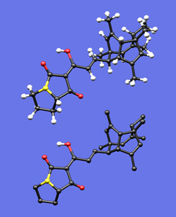
This image was created to accompany an essay I wrote on drawing polycyclic molecules. First I built a plastic model of the molecule and tried to photograph it, but when my photos came out blurry I thought I'd see if I could create an image with my Mac that actually looked like my plastic model. I built the molecule in a molecular modeling program, did a simple force-field minimization, took the resulting coordinates into SwissPdbViewer, and played with the settings (relative radii of balls and sticks, colors, bond lengths) until I felt the feel of my plastic model was captured in the rendered image. I was actually quite surprised how well this worked out -- so much so that someday I may write something on the benefits and dangers of this sort of representation. The bottom image is the same as the top, but with most of the hydrogen atoms removed.
Conservation
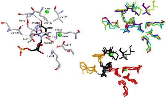
This image was created for a Highlight in ChemBioChem on the crystal structures and mechanisms of the world's most proficient enzyme: orotidine-5'-monophosphate decarboxylase (ODCase). The left panel shows a view of a typical ODCase active site with an inhibitor bound. The top right panel shows a superposition of the active sites from four independently derived x-ray crystal structures of ODCase•inhibitor complexes from four different species (each in a different color). The bottom right panel shows the same superposition now color-coded by residue-type: hydrophobic in black, polar but neutral in gold, charged in red, and waters in green. The molecular structures were visualized using RasMol.
Demoniac Intervention
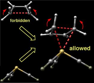
This image depicts the transition state for the disrotatory ring-opening of cyclobutene complexed to iron tricarbonyl. The disrotatory process in the absence of the organometallic fragment is symmetry forbidden, but upon complexation becomes the favored pathway. Woodward and Hoffmann once wrote:
"A very powerful Maxwell demon... could seize a molecule of cyclobutene at its methylene groups, and tear it open in a disrotatory fashion to obtain butadiene. But without such demoniac intervention, molecules will very rarely comport themselves in like fashion." (R. B. Woodward & Roald Hoffmann, Conservation of Orbital Symmetry, 1970)
In a sense, the iron tricarbonyl group here is providing just this sort of demoniac intervention. The molecular structures were visualized with SwissPdbViewer from computationally derived atomic coordinates.
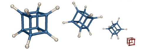
The Same or Not the Same?
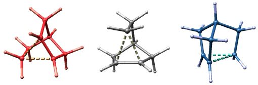
The name of this image alludes to the title of a book written by my postdoctoral advisor (Roald Hoffmann, The Same and Not the Same, 1997). I leave it to you to figure out what these three structures represent. The molecular structures were visualized with SwissPdbViewer from computationally derived atomic coordinates.
In Diamond
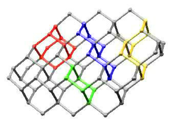
This image highlights chair cyclohexane, adamantane, trans-decalin, and cis-decalin substructures in a portion of the diamond lattice. It is based on an idea I had during graduate school for the cover of an undergraduate organic chemistry textbook. Maybe someday...
Tenuous
The molecule in this image (one designed by Roald Hoffmann years ago) contains two potentially aromatic systems (cyclopropenyl cation and cyclopentadienyl anion) connected by a methylene bridge. Computations may someday address whether these two care to conjoin through a covalent bond or prefer instead to remain in relative isolation. The molecular structure was previsualized with SwissPdbViewer from computationally derived atomic coordinates.
Unbuildable
On occasion, despite the best efforts of authors, editors, and reviewers, a wonderful accident occurs and a drawing of an impossible (or at least highly improbable) molecular conformation slips into the primary research literature of organic chemistry. One such example is shown in this image. The inspiration for creating this image came from Ken Houk, who described these structures as "Escher molecules." The molecular structure in the foreground was previsualized with SwissPdbViewer from computationally derived atomic coordinates.
Cubain't
This image is my spin on the "impossible box" concept (an example is shown in red). The molecular structure of cubane was previsualized with SwissPdbViewer from computationally derived atomic coordinates.
Fickled
This image was created to accompany a paper describing computations on molecules called "fickle hexadienes" and appeared on the cover of the March 8, 2002 issue of the Journal of Organic Chemistry. These molecules can rearrange in two stereochemically distinct ways (one chairlike and the other boatlike), but since the energies of the transition structures for both pathways are the same, the fickle hexadienes have no clear preference for one pathway over the other. The background of the image includes line drawings of the species involved in the rearrangement reactions, and the foreground consists of the two competing transition structures above a stylized potential energy surface adorned with a verse from Shakespeare. The transition state structures were visualized with SwissPdbViewer from computationally derived atomic coordinates. This image was also chosen to appear in the Journal of Organic Chemistry 2003 Calendar.
Models
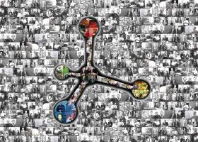
This image of a ball and stick model of a tetrasubstituted methane is composed entirely of images from my collection of pictures of chemists posing with molecular models. It was inspired by a poster I saw in Mike Smith's office at UConn. Click on the image to zoom in.
Oh, Protonation
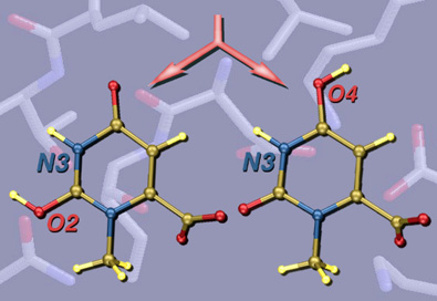
This image was created for Jeehiun Lee. Overlayed on a background of key residues from the active site of the enzyme ODCase are structures of differently protonated substrate models. The active site image was created from x-ray derived coordinates using RasMol, and the protonated substrate structures were created from computationally derived atomic coordinates using SwissPdbViewer.
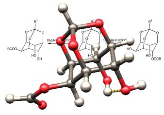
Decarboxylation Machine
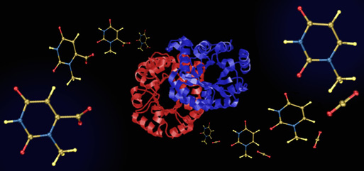
This image was also created for Jeehiun Lee. It consists of a central image of the enzyme ODCase flanked by cascading N-methylorotate molecules on the left and N-methyluracil and carbon dioxide molecules on the right - simple models of the natural substrate for ODCase (orotidine monophosphate) and the products of its decarboxylation. The enzyme image was created from x-ray derived atomic coordinates using RasMol, and the substrate and product structures were created from computationally derived atomic coordinates using SwissPdbViewer. This image appeared in the October 8, 2002 issue of Access. It is also included in the NSF Image Library.
U Exploded
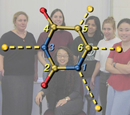
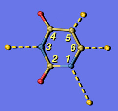
These are two versions of another image created for Jeehiun Lee. Each shows a uracil molecule with its four protons exploded off of the framework. Protons that are farther from the ring are more acidic than those closer to the ring. The background for the version on the left is a photo of the Lee group. The uracil structure was previsualized with SwissPdbViewer using computationally derived atomic coordinates.
Snakes & Ladders
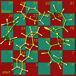
This image shows my version of the classic children's game Snakes and Ladders, using [1,7]-sigmatropic shiftamer transition structures as the snakes and [3,3]-sigmatropic shiftamer transition structures as the ladders. The structures were previsualized with SwissPdbViewer using computationally derived atomic coordinates.
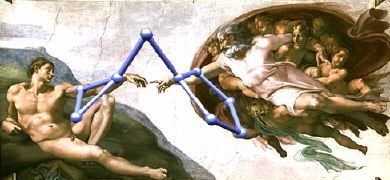
The Circe Effect
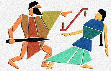
One day I was telling a friend about the enzyme ODCase. She thought I had made up the name as a play on "Odysseus." I thought that was rather funny, especially since one proposed mechanism for ODCase is based on something called the "Circe Effect." In short, this mechanism involves rate acceleration caused by selective destabilization of the reactant when complexed to the enzyme, rather than selective stabilization of the transition state. These two scenarios are shown schematically in red, with Circe favoring reactant destabilization and Odysseus favoring transition state stabilization. Perhaps you can guess which proposal I favor? The layout of this image was inspired by the ancient piece of art shown here.
Full Circle
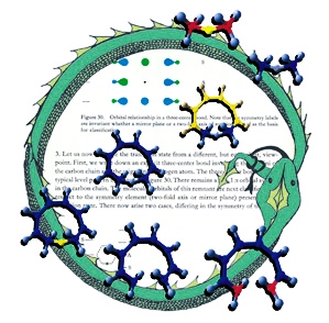
This image was created for the cover of an issue of Angewandte Chemie that featured an article on the geometric and electronic connections between different types of pericyclic reactions. The background is a page from Woodward and Hoffmann's classic Conservation of Orbital Symmetry. The molecular structures are transition structures for [1,8]-sigmatropic hydrogen shifts (center), 8-pi electrocyclization (bottom) and hydride transfer between carbenium ions (top); these structures were previsualized with SwissPdbViewer using computationally derived atomic coordinates. The circle is an orobouros - a mythical serpent eating its own tail - that I created in response to a suggestion by Roald Hoffmann; it is meant to emphasize both the cyclic nature of pericyclic transition structures as well as the fact that the article accompanying this illustration marked Roald's return to the topic of simple pericyclic reactions of hydrocarbons, nearly three decades after he first described many of their key electronic features.
Frame Shifting
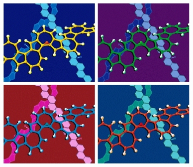
Each frame of this image shows a ball-and-stick model of a transition structure for the shift of a hydrogen atom over one face of a polyazulene polymer in front of line drawings of mono-hydrogenated polyphenacene and polypentalene polymers. A simplified version of this image appeared on the cover of an issue of the European Journal of Organic Chemistry that featured an article on sigmatropic shiftamers. The arrangement of the four frames was obviously inspired by some of the works of Andy Warhol. The structures were previsualized with SwissPdbViewer using computationally derived atomic coordinates.
Insensitivity
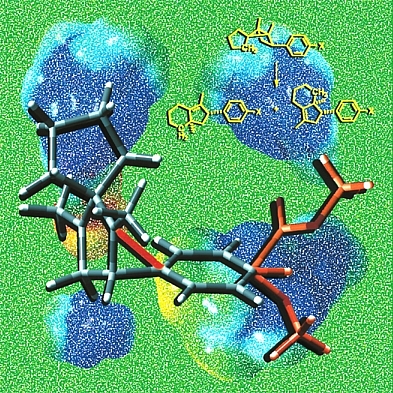
This image shows a model of a 1,3-diyl radical cation, an intermediate in the yellow reaction, stabilized by bridging of an appended aromatic ring. Several different substituents X are shown in orange at the bottom right of the image. Surprisingly, as demonstrated by our collaborator Dan Little, the regioselectivity of the reaction shown is actually insensitive to the nature of the substituent. The background images are based on computed electrostatic potential surfaces of 1,3-diyl radical cations involved in various ring-opening reactions of the type shown in yellow. Structures were previsualized with Gaussview using computationally derived atomic coordinates.
Disconnected
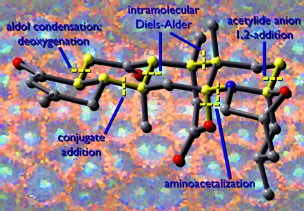
This image shows a model of a complex natural product called norzoanthamine. Synthetic disconnections are highlighted in yellow. The background image is derived from a photograph of the sea anemone (Zoanthus sp.) from which this molecule was first isolated. This image was created for Michael Nantz and George Zweifel based on an idea they had for the cover of their upcoming textbook. The structure was previsualized with Gaussview using computationally derived atomic coordinates.
